Urinary continence is one of the most critical determinants of quality of life following radical prostatectomy. While oncologic cure remains the primary objective of cancer surgery, the fear of permanent pad dependency or involuntary leakage during coughing, laughing, and physical exertion causes understandable anxiety for men diagnosed with localized prostate cancer.
Historically, open radical prostatectomy often treated the pelvic floor as an operative corridor rather than a dynamic suspension apparatus. Dividing the puboprostatic ligaments, incising the endopelvic fascia, and transecting the urethra frequently led to posterior sagging of the bladder neck, shortening of the functional urethral sphincter, and protracted urinary incontinence requiring months or even years of recovery.
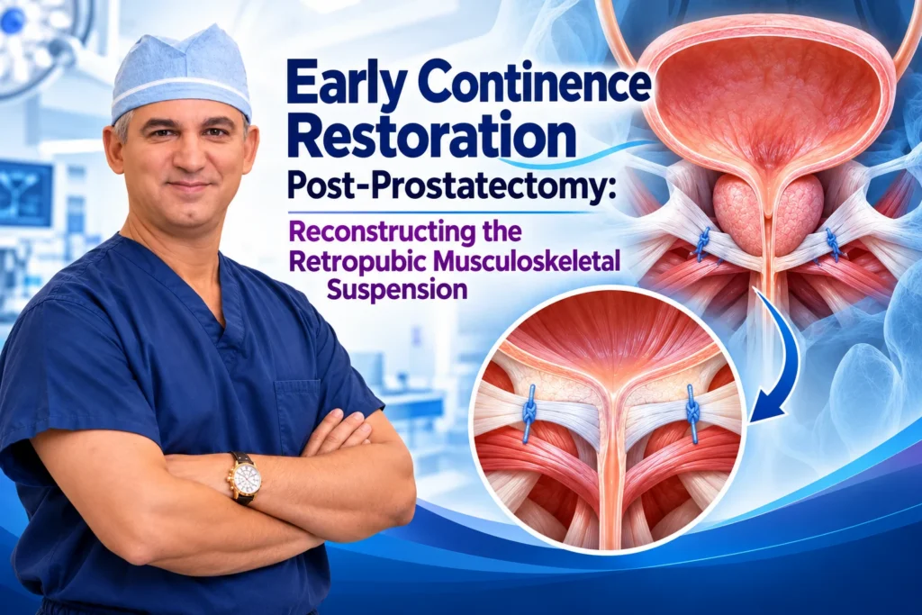
Modern robotic surgery has transformed this outcome. Practicing in New York, Dr. David B. Samadi developed the SMART (Samadi Modified Advanced Robotic Technique) protocol, incorporating advanced anatomical principles to achieve rapid, early continence recovery. By preserving the natural anterior supportive ligaments, maximizing functional urethral length, and reconstructing the posterior musculofascial plate, Dr. Samadi restores the pelvic floor suspension mechanism, allowing the vast majority of patients to regain urinary control shortly after catheter removal.
The Mechanism of Male Continence: The Dual Sphincter System
Normal male urinary continence relies on two integrated sphincteric systems working together:
- Internal Urethral Sphincter (Involuntary): Located at the bladder neck and proximal prostatic urethra, this smooth muscle ring maintains continence at rest under autonomic sympathetic control. Radical prostatectomy inherently removes this internal mechanism along with the prostate gland.
- External Urethral Sphincter (Rhabdosphincter): Located at the membranous urethra just below the prostatic apex, this omega-shaped complex of striated muscle fibers provides active, voluntary control and secondary baseline closure.
Following surgery, the patient must rely entirely on this remaining external rhabdosphincter. If this delicate muscular sleeve is excessively shortened, devascularized, denervated, or left without structural support, post-prostatectomy stress urinary incontinence (SUI) inevitably follows.
The Structural Defect: Why Traditional Surgery Causes Incontinence
When the prostate is excised, the structural connection between the bladder neck and the pelvic diaphragm is interrupted. In conventional prostatectomy approaches, this leads to three primary anatomical disruptions:
- Loss of Urethral Sphincter Length: The membranous urethra is surrounded by the intrinsic sphincter. Aggressive or poorly visualized apical dissection cuts the urethra flush with the pelvic floor, sacrificing several millimeters of functional muscle that are crucial for coaptation.
- Posterior Sagging and Hypermobility: Denonvilliers’ fascia and the retro-urethral connective tissue plate are divided to mobilize the prostate. Without posterior support, the newly created bladder-urethral junction sags downward into the pelvic hollow, creating an abnormal angle that promotes stress-induced leakage.
- Anterior Suspension Disruption: Routine incision of the endopelvic fascia and detachment of the puboprostatic ligaments destabilizes the anterior hammock that holds the urethra upright against the pubic bone during abdominal pressure spikes.
The SMART Suspension Reconstruction Protocol
Dr. Samadi’s continence restoration protocol addresses each of these anatomical challenges through a structured sequence of preservation and reconstruction:
1. Anterior Suspension Preservation
Rather than widely incising the endopelvic fascia and dividing the puboprostatic ligaments—which is standard in many traditional approaches—Dr. Samadi leaves these fibrous bands intact whenever oncologically safe. Sparing the puboprostatic collar keeps the membranous urethra stabilized against the posterior surface of the pubic bone, providing a firm backstop that prevents urethral hypermobility when intra-abdominal pressure rises.
2. Maximum Functional Urethral Length Preservation (MULP)
Under 10x to 15x robotic magnification, the dorsal venous complex (DVC) is divided with cold micro-shears without thermal cautery or bulky bunching sutures. This provides direct visualization of the exact junction between the prostatic apex and the membranous urethra, allowing the surgeon to preserve maximal functional urethral length—typically 15 to 20 millimeters of healthy, vascularized sphincteric tissue.
3. Posterior Musculofascial Plate Reconstruction (Rocco Stitch Variant)
Before fashioning the vesicourethral anastomosis, the retro-urethral supportive floor is restored. Absorbable sutures re-approximate the posterior fibrous layer of Denonvilliers’ fascia to the posterior lip of the rhabdosphincter and the median fibrous raphe. This critical step pulls the urethral stump cranial-ward, eliminates gravitational tension on the healing anastomosis, and reconstitutes the physiological muscular floor.
4. Watertight Running Vesicourethral Anastomosis
A continuous running micro-suture connects the reconstructed bladder neck to the preserved urethral stump. Because tension has already been relieved by the posterior suspension reconstruction, the edges come together naturally without strain. This creates an immediate, watertight seal that minimizes urine extravasation, reduces postoperative inflammation, and facilitates early catheter removal.

Comparative Overview: Conventional Approach vs. SMART Musculoskeletal Reconstruction
| Operative Dimension | Conventional Radical Prostatectomy | SMART Reconstruction Protocol |
| Endopelvic Fascia | Widely incised bilaterally | Preserved intact to maintain anterior suspension |
| Puboprostatic Ligaments | Transected to access dorsal vein | Preserved intact as a structural pubic backstop |
| Apical Dissection | Cautery and pre-emptive bunching suture | Cold micro-dissection under direct visual control |
| Sphincter Length Preserved | Often $< 10\text{ to }12\text{ mm}$ | Maximized ($> 15\text{ to }20\text{ mm}$ of functional urethra) |
| Posterior Pelvic Support | Left open; bladder neck sags into pelvis | Formal re-anchoring of Denonvilliers’ fascia |
| Catheter Removal Timing | Typically 10 to 14 days | Typically 5 to 7 days post-surgery |
| Continence Recovery Profile | 50%–60% at 3 months; gradual over 1 year | Accelerated; high early continence rates |
Postoperative Rehabilitation and Bladder Retraining
Surgical reconstruction provides the anatomical foundation, which is paired with active rehabilitation to support early continence recovery:
- Early Catheter Removal: Because the anastomosis is watertight and tension-free, the transurethral catheter is routinely removed within 5 to 7 days following an in-office evaluation, reducing bladder spasms and urethral irritation.
- Pre- and Postoperative Pelvic Floor Physical Therapy: Patients are instructed in targeted pelvic floor exercises (Kegels) to strengthen the levator ani and pubococcygeus muscles, reinforcing conscious sphincteric control.
- Management of Transient Urgency: Temporary bladder irritability resulting from catheterization is managed with temporary anticholinergics or beta-3 adrenergic agonists if needed, preventing urge-related leakage while the bladder adapts to its new anatomy.
Conclusion
Urinary incontinence does not have to be an inevitable consequence of prostate cancer surgery. By preserving native supportive ligaments, maximizing functional urethral length, and reconstructing the posterior musculoskeletal suspension, the SMART protocol provides patients with an operative pathway designed to eradicate cancer while safeguarding natural urinary control.
- Surgeon: Dr. David B. Samadi, MD
- Specialization: Robotic Urologic Oncology, Prostate Cancer Surgery, Pelvic Reconstruction
- Key Innovations: Developer of the SMART (Samadi Modified Advanced Robotic Technique) Protocol
- Practice Locations:
- Midtown Manhattan: 485 Madison Avenue, 21st Floor, New York, NY 10022
- Long Island: St. Francis Hospital & Heart Center / 2200 Northern Blvd., Suite 120, East Hills, Roslyn, NY 11548
- Official Website:roboticoncology.com
- Direct Consultations: (212) 365-5000
 212-365-5000