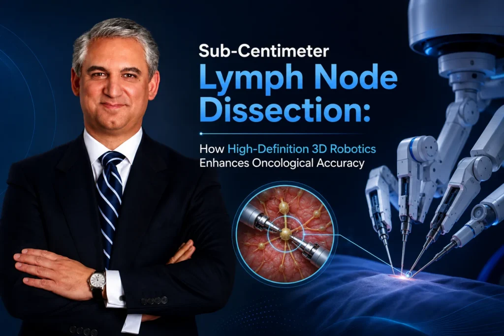
In intermediate- and high-risk prostate cancer, removing the primary tumor inside the prostate gland represents only half of the oncological objective. Because prostate cancer spreads preferentially through the pelvic lymphatic drainage system, microscopic tumor cells often escape into regional pelvic lymph nodes long before they form visible masses on traditional imaging.
Identifying and removing these sub-centimeter lymphatic micro-metastases—nodes that measure under 10 millimeters and frequently go undetected on standard CT or MRI scans—is critical for achieving accurate pathological staging and preventing disease recurrence. However, performing a thorough Extended Pelvic Lymph Node Dissection (ePLND) carries significant surgical complexity, requiring delicate maneuvering around major iliac blood vessels and pelvic nerves.
As Director of Men’s Health and Urologic Oncology at St. Francis Hospital & Heart Center in Roslyn, New York, Dr. David Samadi incorporates ultra-precise, robotic-assisted ePLND into his surgical practice. Backed by his “Samadi Guarantee”—personally performing 100% of every operation—Dr. Samadi leverages 10x 3D optical magnification and tremor-free robotic micro-articulation to perform complete sub-centimeter lymph node dissections without compromising vascular or neural safety.
The Hidden Threat: Sub-Centimeter Nodal Micro-Metastases
Standard cross-sectional imaging (CT and MRI) relies primarily on physical size criteria to identify cancerous lymph nodes. A node is typically flagged as suspicious only when it swells beyond 10 millimeters.
In early lymphatic dissemination, this threshold creates a dangerous diagnostic blind spot:
[Primary Prostate Tumor] ──► Microscopic Cell Shedding into Lymphatic Channels
│
▼
[Sub-Centimeter Pelvic Nodes (<10mm)] ──► Normal size on standard CT/MRI (False Negative)
│
▼
[Extended Robotic Dissection (ePLND)] ──► Physical extraction & true pathological verification
- The Size Illusion: Over 70% of positive pelvic lymph nodes in early-stage prostate cancer measure under 5 millimeters—far below the detection resolution of standard CT scans.
- Pathological Accuracy: Complete surgical excision and microscopic serial sectioning by a surgical pathologist remains the only reliable method to confirm zero nodal involvement (N0 status).
- Therapeutic Benefit: Removing isolated lymphatic micro-metastases prevents those cancer cells from seeding secondary systemic metastases, improving long-term cancer-free survival.
Extending the Dissection Template: Standard vs. Extended Dissection
Surgical precision depends heavily on the anatomical scope of the lymph node removal. A limited dissection leaves crucial lymphatic drainage pathways unexamined.
[Limited Nodal Template] ──► Obstructive zone only (3–5 nodes) ──► Misses high-risk obturator & iliac nodes
VS.
[Extended ePLND Template] ──► Obturator, External Iliac, Internal Iliac, & Presacral zones (15+ nodes)
Dr. Samadi utilizes an extended anatomical template, clearing nodes across all primary pelvic pathways:
- Obturator Fossa Zone: Nodes surrounding the obturator nerve and vessels.
- External Iliac Chain: Lymphatic tissue running along the primary external iliac artery and vein.
- Internal Iliac (Hypogastric) Zone: Deep pelvic nodes adjacent to the internal iliac vessels—a primary drainage site frequently missed in limited dissections.
- Presacral & Common Iliac Packet: Extended clearing up to the bifurcation of the aorta for high-risk clinical presentations.

Standard vs. Extended Pelvic Lymph Node Dissection (ePLND)
| Clinical Parameter | Limited Lymph Node Dissection | Dr. Samadi Extended Robotic ePLND |
| Anatomical Boundary | Restricted to obturator fossa | Obturator, External, Internal Iliac, & Presacral zones |
| Average Node Yield | 4 to 8 nodes | 15 to 25+ nodes retrieved |
| Micro-Metastasis Catch Rate | Misses up to 50% of positive nodes | Highest diagnostic accuracy for sub-centimeter tumor cells |
| Vascular & Nerve Safety | Higher risk of vessel traction in open surgery | Sub-millimeter robotic precision near major iliac vessels |
| Complication Profile | Increased risk of lymphocele if un-sealed | Precision bipolar vessel sealing minimizes fluid accumulation |
How 3D High-Definition Robotics Enhances Safety and Precision
Operating within the deep pelvis to strip microscopic lymphatic packets off thin-walled iliac veins and obturator nerves demands surgical precision. Dr. David Samadi’s proprietary SMART (Samadi Modified Advanced Robotic Technique) procedure delivers key technical advantages during ePLND:
- 10x 3D High-Definition Visualization: Provides crystal-clear visual enlargement of delicate pelvic structures. Dr. Samadi can clearly distinguish between translucent lymphatic channels, tiny micro-vessels, and pelvic nerve fibers.
- Endowrist Instrument Articulation: Robotic micro-instruments offer 7 degrees of freedom, allowing Dr. Samadi to carefully peel sub-centimeter nodal packets off the iliac blood vessels at precise angles that human hands cannot reach through an open incision.
- Athermal & Vessel-Sealing Precision: Advanced energy devices seal lymphatic channels cleanly as they are divided, drastically reducing the post-operative risk of lymphocele (symptomatic fluid collection in the pelvis).
Conclusion
Achieving total victory over prostate cancer requires accurate staging and meticulous removal of hidden sub-centimeter lymphatic micro-metastases. Through high-definition 3D robotic technology and extended lymph node dissection templates, Dr. David Samadi provides men across Long Island and greater New York with rigorous oncological precision, protecting surrounding neural and vascular structures while securing true cancer-free peace of mind.
CTA:Ensure your prostate cancer treatment plan includes comprehensive staging and master-level surgical expertise. Contact the Samadi Robotics Center today at 212-365-5000 to schedule a consultation at our East Hills office located at 2200 Northern Blvd., Suite 120, Roslyn, NY.
 212-365-5000