The widespread use of multiphasic contrast-enhanced CT scans and abdominal MRIs has led to the routine detection of incidental small renal masses. For decades, standard oncologic management defaulted to radical nephrectomy—complete removal of the affected kidney and surrounding Gerota’s fascia. However, long-term nephrology registries confirmed that radical extirpation for localized tumors often traded oncologic cure for an elevated lifetime risk of de novo chronic kidney disease (CKD), secondary cardiovascular events, and all-cause mortality.
Partial Nephrectomy (Nephron-Sparing Surgery) is the recognized reference standard for managing resectable clinical T1 renal tumors. The primary surgical challenge during partial nephrectomy is balancing tumor excision with bleeding control: the kidney receives approximately 20% to 25% of total cardiac output.
To achieve a bloodless field, surgeons traditionally apply vascular bulldog clamps to the main renal artery, inducing warm ischemia. While this facilitates clear visualization during tumor excision, every minute of warm ischemia deprives healthy functional nephrons of oxygen, risking irreversible ischemic cellular death and acute tubular necrosis.
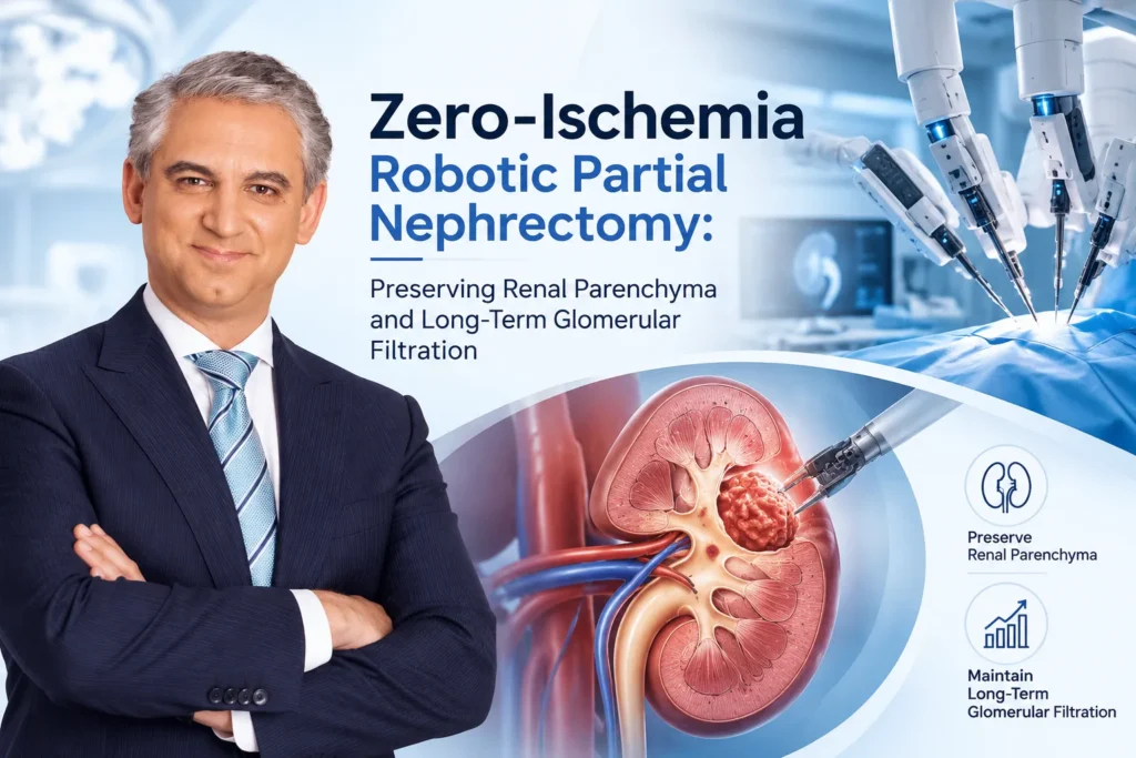
To eliminate this biological insult, advanced urologic oncologists employ Zero-Ischemia Robotic Partial Nephrectomy. By avoiding main renal artery clamping entirely, healthy renal parenchyma remains continuously perfused throughout tumor resection and reconstructive renorrhaphy.
In New York, Dr. David B. Samadi brings extensive background in open oncologic surgery from Memorial Sloan Kettering Cancer Center, advanced laparoscopic fellowship training in France, and thousands of multi-port robotic operations to complex pelvic and retroperitoneal oncology. Utilizing high-magnification 3D optics, micro-dissection, and precise vascular management, Dr. Samadi applies zero-ischemia and super-selective techniques to resect renal masses without compromising global nephron volume or long-term renal function.
The Biology of Renal Ischemia: Why Every Minute Matters
Healthy renal parenchyma contains roughly one million microscopic functional units (nephrons) per kidney. The metabolically active proximal tubular cells within the renal cortex are vulnerable to hypoxia:
[MAIN RENAL ARTERY CLAMPING]
│
▼
[WARM ISCHEMIA CASCADE]
──► Rapid intracellular ATP depletion
──► Cytoskeletal degradation & loss of brush-border membranes
──► Accumulation of reactive oxygen species (ROS)
│
▼
[REPERFUSION INJURY UPON UNCLAMPING]
──► Microvascular vasoconstriction & endothelial cell swelling
──► Influx of inflammatory cytokines (TNF-α, IL-1β)
│
▼
[ACUTE TUBULAR NECROSIS & PERMANENT NEPHRON LOSS]
The Golden Rules of Warm Ischemia Time (WIT):
- $< 20\text{ Minutes}$: Considered acceptable; functional recovery of the remaining kidney depends on baseline reserve.
- $20\text{ to }30\text{ Minutes}$: Measurable subclinical parenchymal injury; elevated risk of acute kidney injury (AKI).
- $> 30\text{ Minutes}$: Significant irreversible ischemic nephron atrophy, focal interstitial fibrosis, and permanent loss of glomerular filtration rate (eGFR).
- Zero Ischemia:Zero warm ischemia time. Capillary blood flow and oxygen delivery remain uninterrupted, eliminating ischemic-reperfusion injury.
Zero-Ischemia Methodologies: How Unclamped Resection is Achieved
Excising a vascularized solid tumor from a fully perfused kidney requires specialized micro-techniques to maintain a dry operative field:
┌──► 1. Anatomical Micro-Dissection (Controlled Hypotension)
│ ──► Resection during controlled systemic mean arterial pressure (MAP 65–70 mmHg)
│
[ZERO-ISCHEMIA TECHNIQUES] ───────┼──► 2. Super-Selective Branch Micro-Clamping
│ ──► Clamping only the 4th/5th-order tertiary feeding arterioles to the tumor
│
└──► 3. Real-Time Near-Infrared Fluorescence (ICG)
──► Firefly™ optical imaging confirming selective tumor ischemia
[Drop-in Robotic Ultrasound Maps Tumor Depth & Endophytic Margins]
│
▼
[Controlled Micro-Dissection along the Fibrous Tumor Pseudocapsule]
│
▼
[Targeted Bipolar Coagulation of Medullary Base Vessels as They Are Encountered]
│
▼
[Frozen Section Histopathologic Margin Verification]
│
▼
[Two-Layer Sliding-Clip Renorrhaphy under Continuous Parenchymal Perfusion]
1. High-Resolution Intraoperative Ultrasound Mapping
Before tumor resection, a flexible drop-in robotic ultrasound probe evaluates the kidney. This defines the tumor’s endophytic depth, locates the proximity of the collecting system (calyces), and maps segmental intrarenal vessels directly underneath the mass.
2. Near-Infrared Fluorescence Imaging (Indocyanine Green – ICG)
Dr. Samadi utilizes intravenous Indocyanine Green (ICG) paired with robotic Firefly™ fluorescence imaging:
- Normal renal cortex expresses high levels of organic anion transporting polypeptides, absorbing ICG and glowing green under near-infrared light.
- Malignant renal cell carcinomas (RCC) lack these transporters and appear dark.
- If super-selective clamping is performed on a tertiary vessel, the devascularized tumor zone turns dark while the surrounding functional cortex remains bright green, verifying uninterrupted blood flow to healthy tissue.
3. Enucleoresection Along the Tumor Pseudocapsule
Rather than taking a wide, destructive margin of normal kidney parenchyma, the robotic micro-shears dissect along the natural fibrous pseudocapsule surrounding the tumor. This plane minimizes blood loss, ensures clear oncologic margins, and preserves the surrounding rim of functional nephrons.
4. Two-Layer Running Renorrhaphy
Hemostasis is established in a structured, two-layer fashion:
- Inner (Medullary) Layer: Bleeding parenchymal vessels and opened calyces are closed with running 3-0 absorbable micro-sutures.
- Outer (Cortical) Layer: The parenchymal defect is re-approximated using sliding-clip renorrhaphy (Hem-o-lok clips), applying uniform compression to the renal cortex without strangulating surrounding tissue.

Preserving Long-Term Glomerular Filtration: The Renal-Cardiovascular Axis
The clinical value of zero-ischemia surgery centers on preserving long-term glomerular filtration:
[RADICAL NEPHRECTOMY] ──► 50% loss of global nephron mass ──► Rapid eGFR decline (< 60 mL/min/1.73m²)
│
▼
[CHRONIC KIDNEY DISEASE (Stage 3+)]
│
▼
[SECONDARY CARDIOVASCULAR RISKS]
├─► Accelerated arterial calcification
├─► Left ventricular hypertrophy (LVH)
└─► Elevated stroke & myocardial infarction risk
[ZERO-ISCHEMIA PARTIAL] ──► Continuous tissue perfusion ──► > 90%–95% baseline eGFR preserved
│
▼
[CARDIOVASCULAR PROTECTION]
(Maintains baseline renal reserve)
Preserving functional parenchymal volume protects patients from subclinical cardiovascular strain, an essential consideration given that most small renal masses present in adults over age 50 who may have coexisting hypertension or diabetes.
Comparative Overview: Warm Ischemia vs. Cold Ischemia vs. Zero Ischemia
| Surgical Metric | Standard Warm Ischemia (Main Artery Clamped) | Cold Ischemia (Ice Slush / Open Flank) | Zero-Ischemia Robotic Protocol |
| Warm Ischemia Time (WIT) | 15 to 25+ minutes | 0 min warm (30–45 min cold) | 0 Minutes (Zero warm ischemia) |
| Parenchymal Perfusion | Completely halted during resection | Completely halted (cooled) | Continuously perfused throughout |
| Reperfusion Injury | Present; microvascular stun | Moderate | None (Zero ischemic insult) |
| Surgical Invasiveness | 3 to 4 robotic keyhole ports | Large, muscle-cutting flank incision | 3 to 4 robotic keyhole ports |
| Postoperative eGFR | Transient drop, gradual recovery | Variable | Preserved; minimal baseline variance |
| Blood Loss (EBL) | Very low ($< 50\text{ mL}$) | Low to moderate ($100–200\text{ mL}$) | Controlled ($< 100–150\text{ mL}$) |
| Hospital Stay | 1 to 2 days | 3 to 5 days | 1 to 2 days (Rapid mobilization) |
Patient Candidacy: Who Benefits from Zero-Ischemia Partial Nephrectomy?
While small, peripheral (exophytic) tumors are primary candidates, zero-ischemia techniques offer meaningful clinical benefits for specific patient populations:
- Pre-Existing Chronic Kidney Disease (Baseline eGFR $< 60\text{ mL/min}$): Patients with diabetic nephropathy or hypertensive nephrosclerosis where any ischemic insult risks precipitating dialysis.
- Solitary Kidney Anatomy: Patients who have previously undergone a contralateral nephrectomy and depend entirely on the remaining kidney for survival.
- Bilateral or Multifocal Renal Tumors: Hereditary kidney cancer syndromes (such as Von Hippel-Lindau disease) requiring sequential partial resections throughout life.
- Complex Polar and Endophytic Lesions: Moderately complex lesions (R.E.N.A.L. Nephrometry score 7–9) where parenchymal volume preservation is balanced with oncologic clearance.
Conclusion
A diagnosis of a solid renal mass does not require sacrificing an entire kidney or exposing healthy nephrons to ischemic injury. Through zero-ischemia robotic partial nephrectomy, intraoperative drop-in ultrasound mapping, and capsule-sparing enucleoresection, Dr. David B. Samadi provides patients with targeted oncologic clearance while safeguarding long-term renal function and cardiovascular health.
- Surgeon: Dr. David B. Samadi, MD
- Specialization: Urologic Oncology, Robotic Surgery, Prostate & Kidney Cancer Specialist
- Key Innovations: Developer of the SMART (Samadi Modified Advanced Robotic Technique) Protocol
- Practice Locations:
- Midtown Manhattan: 485 Madison Avenue, 21st Floor, New York, NY 10022
- Long Island: St. Francis Hospital & Heart Center / 2200 Northern Blvd., Suite 120, East Hills, Roslyn, NY 11548
- Official Website:roboticoncology.com
- Direct Consultations: (212) 365-5000
 212-365-5000