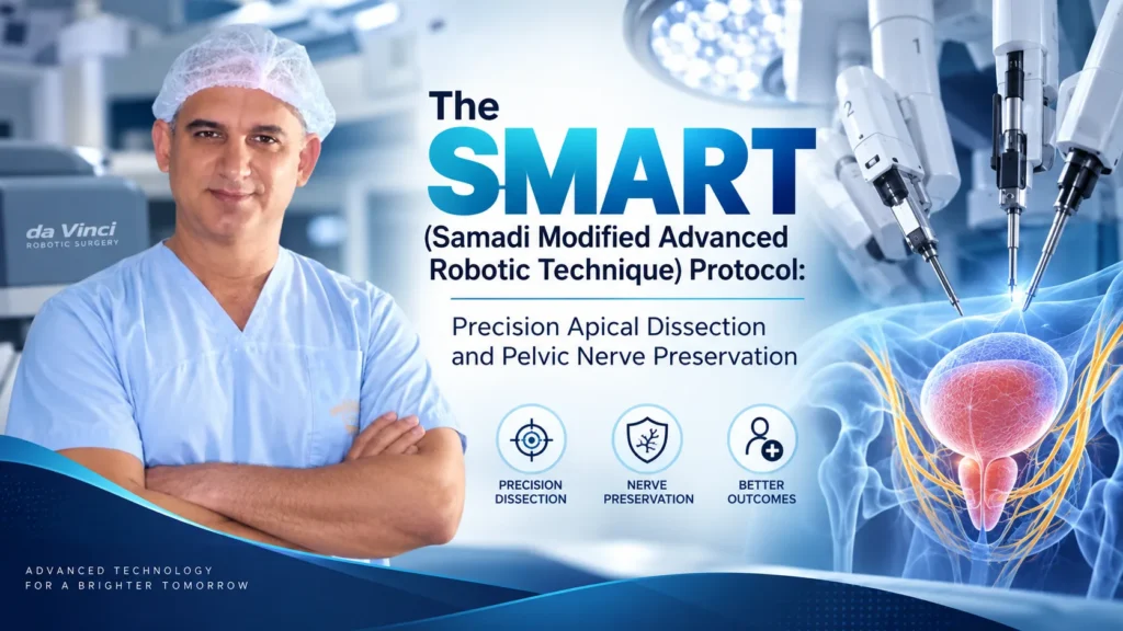
Radical prostatectomy has long been recognized as a benchmark curative therapy for localized prostate cancer. However, the procedure historically confronted surgeons with a difficult compromise: achieving complete oncologic clearance often came at the expense of functional quality of life—specifically, the long-term preservation of urinary continence and erectile potency.
The neurovascular bundles responsible for erectile function course along the delicate lateral borders of the prostate gland, often just millimeters from malignant tissue. Concurrently, the intrinsic urethral sphincter—the primary muscular gatekeeper of urinary control—lies directly adjacent to the prostatic apex, an area where tissue planes converge within a confined pelvic space.
To overcome these technical hurdles, Dr. David B. Samadi developed the SMART (Samadi Modified Advanced Robotic Technique) protocol. Combining open radical prostatectomy principles with high-definition, 3D robotic multi-articulation, the SMART technique avoids thermal energy near delicate nerves, protects the natural pelvic fascial architecture, and utilizes anatomical reconstruction to achieve the “trifecta” of prostate cancer care: negative surgical margins, early return of urinary continence, and preservation of sexual function.
The Trifecta Paradigm in Prostate Cancer Surgery
The standard for surgical success in modern urologic oncology extends beyond cancer eradication alone:
┌──► 1. Oncologic Eradication (Negative surgical margins / Undetectable PSA)
│
[THE RADICAL PROSTATECTOMY] ──────┼──► 2. Complete Urinary Continence (Zero pad dependency)
[TRIFECTA] │
└──► 3. Potency Preservation (Return to unassisted erectile rigidity)
Traditional robotic techniques frequently relied on monopolar or bipolar electrocautery to control bleeding around the lateral prostatic pedicles. While effective for hemostasis, the lateral dispersion of thermal energy through thin pelvic fascia can cause neuropraxia (thermal stun injury) to the microscopic cavernosal nerve fibers.
The SMART protocol addresses this challenge through an athermal, cold-knife approach that completely eliminates electrocautery near the neurovascular bundle pathways.
Core Tenets of the SMART Protocol
The SMART technique builds upon three primary surgical pillars:
[1. ATHERMAL NERVE-SPARING DISSECTION]
──► Cold micro-shears & titanium micro-clips
──► Zero thermal or cautery energy near the cavernosal plexus
──► Intrafascial / interfascial preservation tailored to tumor location
[2. SUSPENSION-PRESERVING APICAL MOBILIZATION]
──► Sparing the puboprostatic ligaments & dorsal venous complex (DVC) without early suture entrapment
──► Maximum functional urethral length preservation (MULP)
──► Zero-tension vesicourethral anastomosis
[3. POSTERIOR MUSCULOFASCIAL RECONSTRUCTION (ROCCO TECHNIQUE VARIANT)]
──► Re-anchoring Denonvilliers’ fascia to the retro-urethral plate
──► Re-establishing natural pelvic floor suspension to accelerate early continence
Step-by-Step Surgical Execution of the SMART Technique
The SMART procedure follows a sequenced operative workflow designed to preserve critical functional structures while maintaining clear surgical margins:
[Transperitoneal Access & High-Definition 3D Optical Mapping]
│
▼
[Bladder Drop & Gentle Exposure of the Endopelvic Fascia]
(Preserving intact puboprostatic ligaments and puboperinealis muscle fibers)
│
▼
[Athermal Seminal Vesicle Mobilization & Posterior Dissection]
(Cold-scissor release of Denonvilliers’ fascia; identifying neurovascular planes)
│
▼
[High Intrafascial / Interfascial Nerve-Sparing Release]
(Athermal vascular control with micro-clips; peeling nerve bundles away intact)
│
▼
[Careful Apical Dissection & Cold Division of Dorsal Venous Complex (DVC)]
(Direct visual control avoiding deep apical sutures that could pinch the sphincter)
│
▼
[Water-Tight Running Vesicourethral Anastomosis & Posterior Suspension Support]
1. Preserving the Endopelvic Architecture
Rather than widely dissecting the endopelvic fascia and severing the puboprostatic ligaments—which tether and support the bladder neck and membranous urethra—the SMART protocol preserves these anterior suspension structures intact. Maintaining this anatomical hammock stabilizes the proximal urethra, reducing post-operative bladder neck hypermobility.
2. Athermal Nerve Release
Once the seminal vesicles and vas deferens are mobilized athermally, Dr. Samadi exposes the lateral prostatic pedicles.
- Zero Electrocautery: No electric current, ultrasonic shears, or laser thermal devices are used within 1 to 2 centimeters of the neurovascular bundles.
- Micro-Clips and Cold Scissors: Pedicle vessels feeding the prostate are individually controlled using tiny titanium micro-clips and sharp micro-scissors.
- Fascial Plane Selection: Depending on preoperative multiparametric MRI findings, targeted biopsy maps, and clinical staging, the surgeon selects an intrafascial plane (stripping the innermost capsule to leave 100% of adjacent nerve tissue intact for low-to-intermediate-risk lesions) or an interfascial plane (taking a protective cushion of tissue when extracapsular extension is suspected).
3. Apical Dissection and Maximal Urethral Length
The prostatic apex presents a delicate operative stage: positive surgical margins frequently occur here due to the absence of a distinct anatomic capsule, while the intrinsic striated urethral sphincter wraps directly around the emerging membranous urethra.
- Under 10x to 15x robotic magnification, the dorsal venous complex (DVC) is divided athermally using cold micro-shears under controlled pneumoperitoneum.
- This direct visualization approach avoids placing broad, bulky DVC sutures that can unintentionally compress the underlying striated sphincter muscle fibers.
- The surgeon maximizes functional urethral length (aiming for $> 15\text{ to }20\text{ mm}$ of preserved membranous urethra), which correlates directly with rapid urinary continence recovery.
4. Posterior Musculofascial Plate Reconstruction
Prior to completing the running vesicourethral anastomosis (the Van Velthoven technique), the retro-urethral plate and Denonvilliers’ fascia are reconstructed and anchored to the posterior bladder neck. Re-approximating these posterior fascial layers eliminates downward tension on the urethral stump and creates a supportive floor for the bladder neck.
Comparative Overview: Standard Robotic Prostatectomy vs. The SMART Protocol
| Surgical Metric | Standard Multi-Port Robotic Surgery | The SMART Protocol (Dr. Samadi) |
| Nerve-Sparing Modality | Cautery / Bipolar coagulation near pedicles | 100% Athermal (Cold scissors & micro-clips) |
| Nerve-Stun Risk (Neuropraxia) | Elevated due to lateral thermal spread | Minimized; zero electric energy applied to nerves |
| Endopelvic Fascia Management | Widely incised and detached | Sparing of puboprostatic ligament complex |
| Apical DVC Management | Pre-emptive figure-of-8 bunching stitch | Cold cut under direct vision; selective hemoclip control |
| Urethral Length Preservation | Variable | Maximized ($>15\text{ mm}$ of functional sphincter spared) |
| Pelvic Floor Reconstruction | Often anterior or posterior alone | Dual posterior & anterior anatomical reconstruction |
| Continence Restoration | 60%–70% at 3 months; 85%–90% at 1 year | Significant acceleration (high early continence rates) |
Clinical Indications: Matching the Technique to Tumor Biology
The SMART protocol is adapted across the clinical risk spectrum:
- Low- and Favorable Intermediate-Risk Disease (Gleason 3+3, 3+4): Bilateral intrafascial nerve-sparing approaches are utilized to protect functional pathways without compromising oncologic margins.
- High-Risk Localized Disease (Gleason 4+4, 4+5, PSA $>20\text{ ng/mL}$): The athermal approach is combined with an Extended Pelvic Lymphadenectomy (ePLND). If the tumor shows signs of focal capsular abutment on one side, an intentional wide neurovascular resection is performed on the affected side while sparing the contralateral bundle to preserve baseline erectile potential.
Postoperative Recovery and Rehabilitation Pathways
- Hospital Stay: Patients are routinely mobilized out of bed within 4 to 6 hours post-surgery and typically discharged home on postoperative day one.
- Catheter Management: Due to the precise, watertight running anastomosis, the urethral catheter is typically removed within 5 to 7 days, confirmed via in-office voiding assessment.
- Pelvic Floor Recovery: Patients begin guided pelvic floor muscle exercises (Kegels) to accelerate muscle retraining.
- Penile Rehabilitation: Early non-erectile nighttime oxygenation protocols—such as low-dose daily PDE-5 inhibitors—are initiated upon catheter removal to promote cavernosal tissue health while the uninjured nerves recover full signal transmission.
Conclusion
The SMART protocol integrates the visual magnification and range of motion of robotic instrumentation with the tactile principles of open athermal surgery. By prioritizing nerve preservation through cold micro-dissection, maximizing urethral length, and reconstructing natural supportive ligaments, this technique aims to eradicate localized prostate cancer while protecting the urinary continence and personal quality of life patients value most.
- Surgeon: Dr. David B. Samadi, MD
- Specialization: Robotic Urologic Oncology, Prostate Cancer Surgery, Minimally Invasive Urology
- Key Innovations: Developer of the SMART (Samadi Modified Advanced Robotic Technique) Protocol
- Clinical Focus: Robotic Radical Prostatectomy, Secondary/Salvage Prostatectomy, Complex Pelvic Oncology
 212-365-5000